Digital Pathology Defined
We Use Image Recognition & Data Sciences
The video below, produced by the Nasjonal IKT of Norway illustrates
how the digital pathology system works and describes many benefits of digital pathology.
Histofi Digital Pathology Workflow
Histofi digital pathology system is a complete workflow by design
Only digital pathology system integrated with AP-LIS and Quality control
Anatomic Pathology-LIS Integrated:
Digital Pathology
We addressed quality control issues in anatomic pathology labs, which drive quality in digital pathology outcomes. Our digital pathology workflow is a complete suite integrated with the following sub-systems:
- Biopsy sample management (full functionality)
- LabNote with step-wise protocols to ensure quality
- Sample shipment, tracking, and reporting
- Billing and revenue collection
- Molecular pathology module (optional)
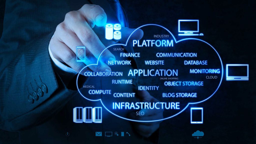
Histofi SaaS Platform
We distribute AI-powered digital pathology solutions to providers anywhere using a SaaS platform. Our platform ensures quality digital pathology workflow leading to faster diagnosis and collaborative treatment decision support.
- Cancer-specific diagnostic AI application
- Telepathology – second opinion
- Tumor board services
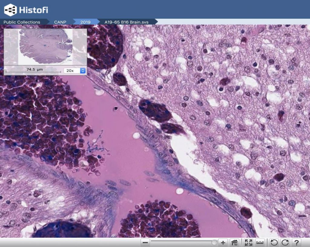
Histofi supports all major digital pathology formats, allowing you to select the scanner that best meets your needs. Our select scanners are fully integrated with Histofi image management and analytic system. Features include:
- Scanner agnostic: Chose your scanner or procure Histofi’s integrate one.
- Zero-footprint: the browser-based interface eliminates the need to download and install client software. View and manage your images using IE, Edge, Chrome, or Firefox on either PC, MAC, or mobile device.
- Organize: Create folders and organize images based on your workflow requirements. Upload images directly to your workspace and have them immediately available for viewing and sharing with colleagues.
- Image analysis: Perform quantitative IHC analysis using Immunopath Image Analysis. Develop protocols or utilize and modify existing protocols for nuclear, cytoplasm, or membrane analysis on WSI, microscopic, or TMA images.
- Annotation and Measurement: Simple and easy to use tools for annotations and measurements. Bookmark your annotations to quickly navigate to important areas of interest, Ideally suited for remote consultations or e-learning applications.
- Share images for a second opinion: encrypted URL link to an image file that can be shared by email or instant message. Collaborators can click on the link and get secure direct access to review the full digital image file. DirectLink™ provides a fast, effective tool for enabling consultations, 2nd opinions, and research collaborations.